<<<
Compare
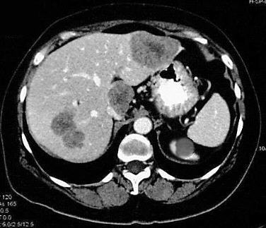
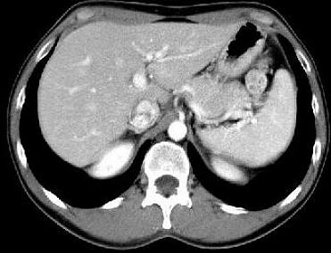
the pathological image-left and the physiological image-right
(blinded)
<<
F:
Multiple
hypodense hepatic structures
with a central dark zone
H:
Adult
woman, 61-years-old, known sigmoid carcinoma
INFO/WWW-LINKS:
Colorectal carcinomas are adenocarcinomas which may often arise in pre-existing
polyps. About 40% of these carcinomas are located in the rectum, 20-30% in
the sigmoid colon, and about 30% in the descending, the transverse and coecum
with ascending colon. Colorectal carcinomas spred local, lymphatic and haematogenous
- especially - to the liver
and the lung.
D:
Multiple
hepatic metastases from a carcinoma of the sigmoid colon.
Cyst
of the left kidney
IN
THIS PART OF THE PAGE YOU FIND SOME TEXT FIELDS WHICH CAN BE OPENED EIGTHER
STEP BY STEP (CLICK ON "HISTORY", "HELP", "FINDINGS",
"DIAGNOSIS" OR "INFO/WWW-LINKS") OR AT ONCE WITH A CLICK
ON "ALL ON" - VICE VERSA CLICK ON "ALL OFF".
It is not
easy to find an exactly corresponding slice to every pathological example!
For that reason the
FILM
(2)
is
recommended!
Once opened you may use it for every pathological example.
If you need a physiological
image to compare click here