<<<
Compare
the pathological image-left and the physiological image-right
(blinded)
<<
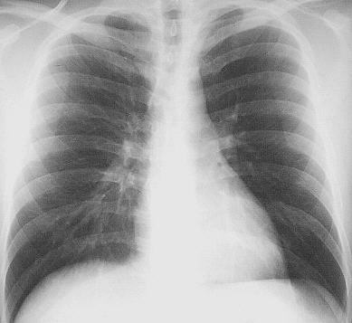
H:
Adult man, 23-years-old, repeated dyspnoe, fever and recurrent chest infections
INFO/WWW:
Bronchogenic csyts
are thin-walled cystic structures, having a lining of respiratory epithelium
and containing mucoid material. Sometimes there is connection to the tracheobronchial
tree. Bronchogenic cysts are usually located near the trachea or main bronchi.
Patients with bronchogenic cysts are ususally asymptomatic. But hemoptysis,
infections, or rupture into the trachea, lung, esophagus or pleural space
may occur; large proximal cysts occasionally compress major airways or blood
vessels.
D:
Right-sided hilar bronchogenic cyst with an air-fluid level as evidence for
infection.
IN
THIS PART OF THE PAGE YOU FIND SOME TEXT FIELDS WHICH CAN BE OPENED EIGTHER
STEP BY STEP (CLICK ON "HISTORY", "HELP", "FINDINGS",
"DIAGNOSIS" OR "INFO/WWW-LINKS") OR AT ONCE WITH A CLICK
ON "ALL ON" - VICE VERSA CLICK ON "ALL OFF".
If you need
a physiological image to compare click here